医療工学
医用画像の視認性向上と診断支援に関する研究
| 特記事項:
本研究は,独立行政法人科学技術振興機構が推進する平成15年度大学発ベンチャー創出事業において,『全方位ビジョンを用いた医療技術の開発』として課題採択されました. |
| Color Analysis for Segmenting Digestive Organs in VCE |
|
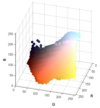 |
This paper presents an efficient method for automatically segmenting the digestive organs in a Video Cap- sule Endoscopy (VCE) sequence. The method is based on unique characteristics of color tones of the digestive organs. We first introduce a color model of the gastroin- testinal (GI) tract containing the color components of GI wall and non-wall regions. Based on the wall re- gions extracted from images, the distribution along the time dimension for each color component is exploited to learn the dominant colors that are candidates for discriminating digestive organs. The strongest candi- dates are then combined to construct a representative signal to detect the boundary of two adjacent regions. The results of experiments are comparable with previ- ous works, but computation cost is more efficient. |
|
|
| カプセル内視鏡を用いた小腸収縮位置の検出 |
|
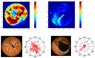 |
This paper describes a method for automatic detection of contractions in the small bowel through analyzing Wireless Capsule Endoscopic images. Based on the characteristics of contraction images, a coherent procedure that includes analyzes of the temporal and spatial features is proposed. For temporal features, the image sequence is examined to detect candidate contractions through the changing number of edges and an evaluation of similarities between the frames of each possible contraction to eliminate cases of low probability. For spatial features, descriptions of the directions at the edge pixels are used to determine contractions utilizing a classification method. The experimental results show the effectiveness of our method that can detect a total of 83% of cases. Thus, this is a feasible method for developing tools to assist in diagnostic procedures in the small bowel. |
|
|
| 医療用内視鏡のための全方位視覚アタッチメント |
|
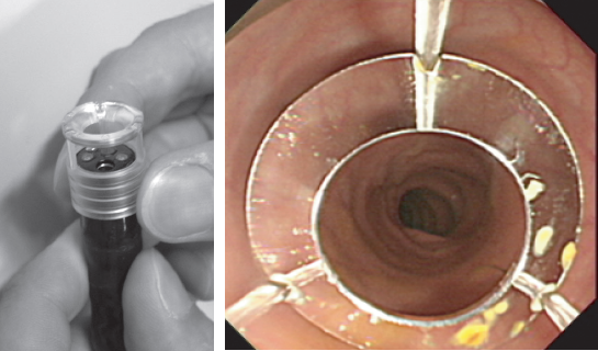 |
大腸など人間の臓器内部は,襞などによって複雑な形状をしているため,内視鏡の後方が死角となり,患部を見落とす可能性が指摘されている.本論文では,前方だけでなく,周囲360度の側方並びに後方観察が可能な全方位消化管内視鏡を提案する.本方式は,通常の消化管内視鏡の先端部に環状凸面鏡の使い捨て型全方位アタッチメントを装着することで,前方だけでなく側後方を同時に観察することができる.内視鏡医による人体モデル並びに動物実験により,本アタッチメントの有用性,安全性を確認した. |
|
|
| カプセル内視鏡映像における適応的表示速度制御方式 |
|
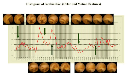 |
カプセル内視鏡で撮影された8時間映像を,病巣を見落とさずに注視し続けるのは医師にとって負担である.現状では,画像描画速度を手動で変更し,早送り・スロー再生によって診断するため,熟練者でも45分以上,初心者では2時間程度の診断時間を要している.そこで,ビデオ表示速度を画像処理によって適応制御し,煩雑な手作業を軽減することで短時間の診断が可能となる支援ツールを実現する.本研究では,連続する画像の特徴量である隣接画像間類似度と移動量から,映像取得時におけるカプセルおよび小腸の状態を分類し,状態によって描画速度を決定する.その状態判定と速度決定には多くのパラメータを含むため,複数の医師の評価から最適なパラメータを決定する.結論として,8時間映像を適応制御による30分程度の表示でも診断可能になり,平均再生速度15倍速を達成した. |
|
|
| 消化器官展開画像生成 |
|
 |
消化器官内視鏡映像による検査では,移動するカメラから撮影した画像列を獲得するが,病変の確認には,画像列を繋ぎ合わせた静止画として描画する方が容易である.本研究では,超広角レンズまたは全方位ミラーから獲得したカメラ側方の環状画像列から,投影面を一般化円筒の制約以外に既定しないビデオモザイキングを行う.環状画像を展開した画像は隣接フレーム間の移動が多項式近似可能で,変形した展開画像を多数フレームで貼り合せることから,腸管展開画像を生成する. |
|
|
| 内視鏡カメラの歪み補正 |
|
 |
消化器官内視鏡は,カメラの周囲映像を捉えるために広角レンズが用いられている.本研究では,広角レンズによるレンズ歪み補正を,平面ディスプレイ上に表示したグレイコードパターンを利用することで,ディスプレイ座標系とカメラ画像座標系の対応関係となる変換マップ作成によって実現する.本手法は,従来手法と異なりレンズ歪みモデルを用いず,ディスプレイと画像の密な対応関係から,画像周辺部でも適応可能であり,単純な線形補間によって元画像から歪み補正画像への変換マップが作成可能となる利点がある.また提案手法は,変換マップを直接作成して歪みを補正するので,画像中心・アスペクト比・光軸からの傾き角のカメラ内部パラメータを必要としないのが特徴である. |
|
|