My research is in the area
of medical image processing and analysis,
focusing on the capsule endoscopic images. We conduct approaches to
assist the capsule endoscopists in reading video sequences as well as understanding the intestinal motility using image sources. Topics include:
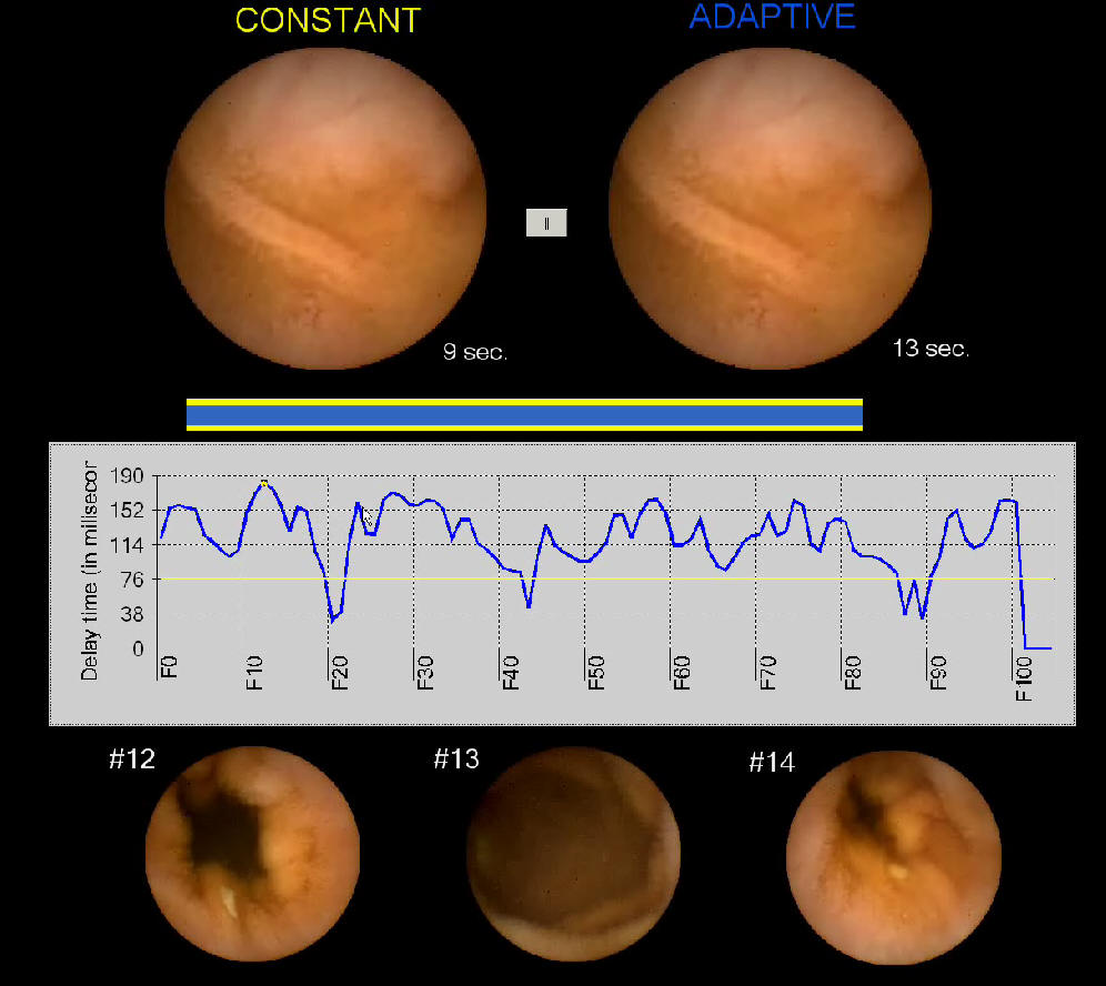
[R1] Reducing Diagnostic Time
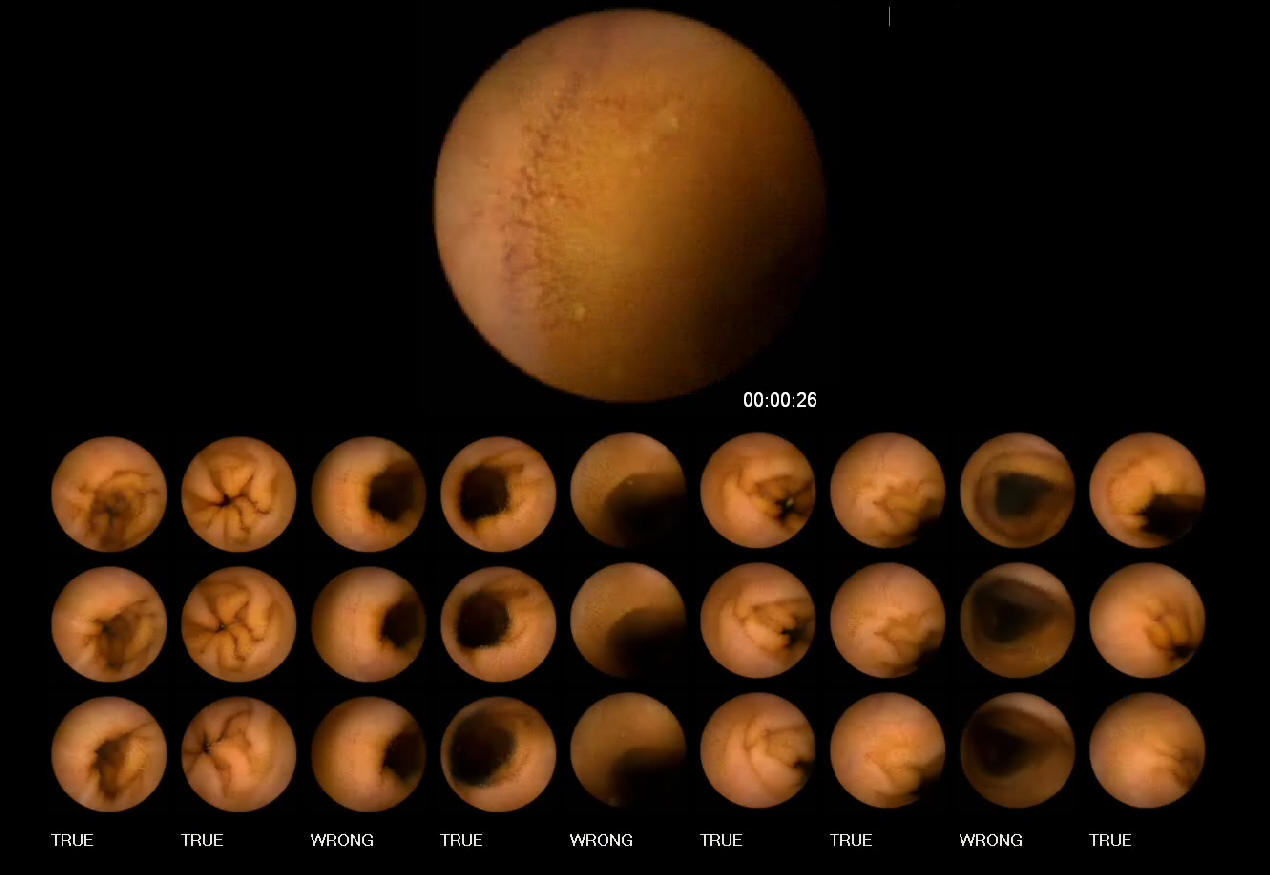
[R2] Intestinal Contraction Detection
[R3] Segmenting Digestive Organs in Video Capsule Endoscopy
[R4] Image-Enhanced Capsule Endoscopy Preserving the Original Color Tones
Image-Enhanced Capsule Endoscopy Preserving the Original Color Tones
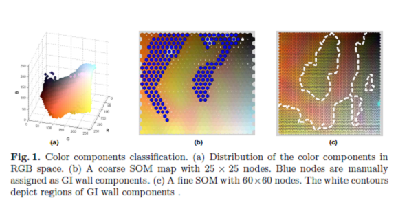
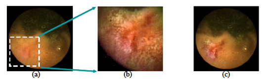
This paper describes a technique to enhance a region-of-interest (ROI) in a capsule endoscopy (CE) image. Our aim is to improve distinguishing suspicious regions from normal ones without over enhancing the image. Given a ROI by physicians, the proposed technique enhances the ROI so that the enhanced region remains as natural as possible. To achieve this, we utilize image features relating to a specific color
space dedicated to gastrointestinal (GI) wall regions. The proposed approach starts by utilizing a self-organizing map to handle GI wall color components. The goal of preserving the original color tones is obtainable by using a histogram equalization technique in the proposed color space. As a result, the enhanced images only contain color components inferred from the color gamut of the human small bowel. The results of the proposed method are judged in terms of visual appearance by comparison
with images obtained by the conventional enhancement technique.
Image Enhancement with user-interactions
Segmenting Digestive Organs in Video Capsule Endoscopy
This paper presents an efficient method for automatiically segmenting the digestive organs in a Video Cap sule Endoscopy (VCE) sequence. The method is based on unique characteristics of color tones of the digestive organs. We first introduce a color model of the gastrointestinal (GI) tract containing the color components of GI wall and non-wall regions. Based on the wall regions extracted from images, the distribution along the time dimension for each color component is exploited to learn the dominant colors that are candidates for discriminating digestive organs. The strongest candidates are then combined to construct a representative signal to detect the boundary of two adjacent regions. The results of experiments are comparable with previous works, but computation cost is more efficient.
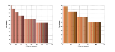
Dominant Colors in Stomach and Intestinal Wall regions
Segmenting Pylorus and IV in a Video Capsule Endoscopy
Display of a capsule video sequence is controlled by a technique known as "Adaptive Display Speed". Experiments conducted from four examining doctors and six sequences showed that average of the diagnostic time is 32.5 min (std=± 7 min.) for an evaluation. (sample_1.avi, sample_2.avi)